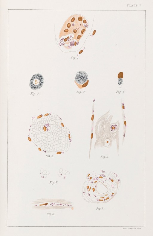
Illustration of cells infected with Leprosy bacilli.
Unknown artist
Plate X Fig 1. Epithelial cell from a seminal canal, filled with bacilli Fig 2. A globus with a vacuole in which are fragments stained with Bismarck brown, probably the remains of nuclei Fig 3. a globus lying in a cell; the nucleus and a part of the cell-protoplasm preserved. Fig 4. An epithelial cell from a seminal canal, filled with bacilli broken down into granules. Fig 5. Cross section of a bloood-vessel with bacilli in the endothelium and a white blood corpuscle filled with bacilli. Fig 6 Longitudinal section of a blood-vessel showing bacilli in the endotheluim and a fibrinous coagulum enclosing two white blood corpuscles, one of them filled with bacilli. Fig 7 Bacilli free between the red blood corpuscles. Fig 8 A connective tissue space from the tunica albuginea (dense covering of testicle) filled with bacilli Fig 9. Cross section of a blood-vessel with bacilli around the nuclei of the surrounding connective tissue. All the preparations from the testicle were hardened in Fleming's or Müller's solutions, and the drawings made with a Seibert Homog. immersion lens 1/16.
Sign in to order prints, canvas, and more of this work. Browsing and full-resolution downloads stay free.


