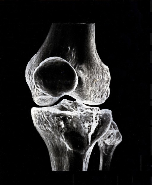
X-ray of a knee joint printed on coated aluminium, artwork
Chiara Salvi
A 3D digital representation of a knee joint Xray, printed onto aluminium plates. The xray image was printed digitally onto acetate and used as a negative. Silver gelatin was used to coat aluminium plates, making them photosensitive. These coated aluminium plates were then treated as a photographic piece of paper: the artist has used the traditional analogue processing methods of developing and fixing to produce the final images. This method was the primary processing method between the 1880s and the 1960s, before colour photographs gained popularity. This image is of the knee joint as seen from the front. The knee cap, or patella, is represented by the circle in the centre of the image. The bone above the joint is the femur, and two bones below are the tibia (left) and fibulla (right). This image is part of a bigger project called 'Soffice' or 'De Humani Corporis Fabrica' The original artwork is 11 x 14 cm.
Sign in to order prints, canvas, and more of this work. Browsing and full-resolution downloads stay free.