Wellcome Collection
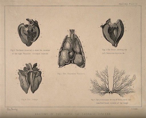
Internal organs and arteries of the horse: five figures showing dissections of the heart, lungs, thoracic viscera, pulmonary artery and veins. Engraving by T. Cowan after B. Herring, ca. 1860.
Benjamin Herring · 1860
Lettering: Anatomy of the horse. The thoracic viscera ; fig. 1, The thoracic viscera ; fig. 2, The heart dissected to show the interior of the right ventricle, tricuspid valve &c. ; fig. 3, The heart, showing the left ventricle, auricle etc. ; fig. 4, The lungs ; Fig. 5. The pulmonary artery & veins, with the ramified blood-vessels of the lungs. Bears number: Plate 12. Figures individually lettered for key.
🔒
Order museum-quality prints
Sign in to order prints, canvas, and more of this work. Browsing and full-resolution downloads stay free.


