Wellcome Collection
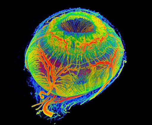
3D view on vessels of a healthy minipig eye. The upper opening corresponds to the pupil as the gateway input of all light into the eye. It is interesting to see the marked abundance of vessels of the pupil which bring energy and food to the muscles to control the amount of incident light. The other large vessels are feeder vessels for the outer layers of the retina and muscles, so, that the eye quickly can perceive the environment and the creature may adapt and survive.
Dr Peter M Maloca
Information about the blood vessels was taken by injection of a newly developed, high-viscous contrast media. Micro-CT scanning was applied and a 3D vessel volume rendered. Images were freed from speckle-noise with a three-dimensional filter to enhance the signal. Obtained vessels were color-coded. The axial length is 19 millimeter. The smallest vessels that have been detected are 20-30 microns in diameter.
🔒
Order museum-quality prints
Sign in to order prints, canvas, and more of this work. Browsing and full-resolution downloads stay free.


